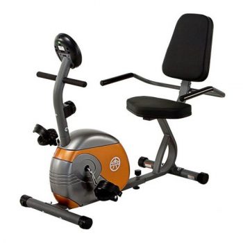
In stock
Mindray M7 Diagnostic Ultrasound System
$299.00
The Mindray M7 epitomizes the optimal combination of advanced imaging technologies and miniaturization technologies. Mindray research and development engineers employ the System On Chip (SOC) design within the M7 ultrasound system. SOC enables complex technologies to be built into the M7’s compact laptop style chassis. Medical devices built around SOC designs are energy efficient and highly reliable. The M7’s exceptional image quality, user experience, and versatility have expanded the performance and flexibility envelope for hand carried ultrasound imaging systems.
-
Guarantee safe checkout
Flexibility to meet your challenging needs today and tomorrow Advanced and versatile technologies that work effectively for multiple specialties • Excellent system performance across a wide range of applications • User selectable setup, from dedicated specialty to full service • Clinical application-specific measurement, calculation and reports • Comprehensive DICOM features: Store/Print, Worklist, MPPS, Query/Retrieve, OBGYN S/R, Vascular S/R and Cardiac S/R • Wired and wireless data transfer and connectivity • System ergonomic and utility: hand-carried to full service cart with external display • Durable chassis, 1.5 meter drop tested, meets the transport and handling demands of various care environments • Upgradability: – Expansion of current system capability – Addition of new capabilities in future releases The Mindray M7 diagnostic ultrasound system: your best choice for quality, speed and flexibility.
Endovaginal scan of the uterus with wide field of view.
Transverse scan of thyroid showing near field detail and high sensitivity color Doppler
Liver imaging with excellent detail and uniformity
Color Doppler image of carotid artery with automatic spectral doppler quantification
Real-time 4D surface rendering of fetal face
FreeXros M, anatomical M mode of mitral valve
IMT, auto-measurement of carotid intimae-thickness
Mitral regurgitation imaged in color Doppler and spectral Doppler modes
High frequency imaging of median nerve with excellent detail and contrast resolution
Imaging with clarity and accuracy: the basis for diagnostic confidence.
Across a wide range of clinical specialties: abdomen, obstetrics, gynecology, cardiology, peripheral vessels, small parts, urology, anesthesia, emergency medicine, ICU/CCU, pediatrics, neonate, transcranial, interventional, musculoskeletal and intra-operative procedures.
Download From Here >>Flexibility to meet your challenging needs today and tomorrow Advanced and versatile technologies that work effectively for multiple specialties • Excellent system performance across a wide range of applications • User selectable setup, from dedicated specialty to full service • Clinical application-specific measurement, calculation and reports • Comprehensive DICOM features: Store/Print, Worklist, MPPS, Query/Retrieve, OBGYN S/R, Vascular S/R and Cardiac S/R • Wired and wireless data transfer and connectivity • System ergonomic and utility: hand-carried to full service cart with external display • Durable chassis, 1.5 meter drop tested, meets the transport and handling demands of various care environments • Upgradability: – Expansion of current system capability – Addition of new capabilities in future releases The Mindray M7 diagnostic ultrasound system: your best choice for quality, speed and flexibility.
Endovaginal scan of the uterus with wide field of view.
Transverse scan of thyroid showing near field detail and high sensitivity color Doppler
Liver imaging with excellent detail and uniformity
Color Doppler image of carotid artery with automatic spectral doppler quantification
Real-time 4D surface rendering of fetal face
FreeXros M, anatomical M mode of mitral valve
IMT, auto-measurement of carotid intimae-thickness
Mitral regurgitation imaged in color Doppler and spectral Doppler modes
High frequency imaging of median nerve with excellent detail and contrast resolution
Imaging with clarity and accuracy: the basis for diagnostic confidence.
Across a wide range of clinical specialties: abdomen, obstetrics, gynecology, cardiology, peripheral vessels, small parts, urology, anesthesia, emergency medicine, ICU/CCU, pediatrics, neonate, transcranial, interventional, musculoskeletal and intra-operative procedures.
Download From Here >>Description
chassis. Medical devices built around SOC designs are energy efficient and highly reliable. The M7’s exceptional image quality, user experience, and versatility have expanded the performance and flexibility envelope for hand carried ultrasound imaging systems.
Equipped for Quality
Radiology
Cardiology
Outpatient
Emergency
Flexibility to meet your challenging needs today and tomorrow
Advanced and versatile technologies that work effectively for
multiple specialties
• Excellent system performance across a wide range of applications
• User selectable setup, from dedicated specialty to full service
• Clinical application-specific measurement, calculation and reports
• Comprehensive DICOM features: Store/Print, Worklist, MPPS, Query/Retrieve, OBGYN S/R, Vascular S/R and Cardiac S/R
• Wired and wireless data transfer and connectivity
• System ergonomic and utility: hand-carried to full service cart with external display
• Durable chassis, 1.5 meter drop tested, meets the transport and handling demands of various care environments
• Upgradability:
– Expansion of current system capability
– Addition of new capabilities in future releases
The Mindray M7 diagnostic ultrasound system: your best choice for quality, speed and flexibility.


Endovaginal scan of the uterus with wide field of view.
Transverse scan of thyroid showing near field detail and high sensitivity color Doppler
Liver imaging with excellent detail and uniformity
Color Doppler image of carotid artery with automatic spectral doppler quantification
Real-time 4D surface rendering of fetal face
FreeXros M, anatomical M mode of mitral valve
IMT, auto-measurement of carotid intimae-thickness
Mitral regurgitation imaged in color Doppler and spectral Doppler modes
High frequency imaging of median nerve with excellent detail and contrast resolution
Imaging with clarity and accuracy: the basis for diagnostic confidence.
Across a wide range of clinical specialties: abdomen, obstetrics, gynecology, cardiology, peripheral
vessels, small parts, urology, anesthesia, emergency medicine, ICU/CCU, pediatrics, neonate, transcranial, interventional, musculoskeletal and intra-operative procedures.


Reviews
There are no reviews yet.